February 21, 2024
Softsmile’s Vision CBCT 3D Imaging Capabilities & Functionality


SoftSmile has spent years developing the Cone Beam Computed Tomography (CBCT) data analysis capabilities of its treatment planning software VISION. An updated version of VISION was launched this past summer with advanced CBCT analysis. VISION’s ability to manipulate complex data from CBCT 3D imaging scans allows for new levels of analysis and diagnosis that improve treatment planning design and outcomes.
Below are important questions and answers about the capabilities and functionality of VISION’s CBCT:
How long does it take to process a scan CBCT and merge it with an intra-oral scan in VISION?
There is no manual effort required by the user. This functionality is automated in VISION and processing takes 2-3 minutes.
What file formats are accepted for VISION’s CBCT?
The industry standard is DICOM (DICOM files contain a patient's x-ray image or series of images and other patient-related information) which VISION accepts as a folder or zip file.
Does VISION accept DICOM files from any CBCT brand?
Yes. The VISION algorithm is trained on a wide variety of settings, and while there may be some files that are outliers, the VISION algorithm will improve over time.
Does VISION accept both hollow and solid model scans?
Yes, VISION accepts both solid and hollow arch scans. For Dental CBCT scans, VISION requires volumetric data.
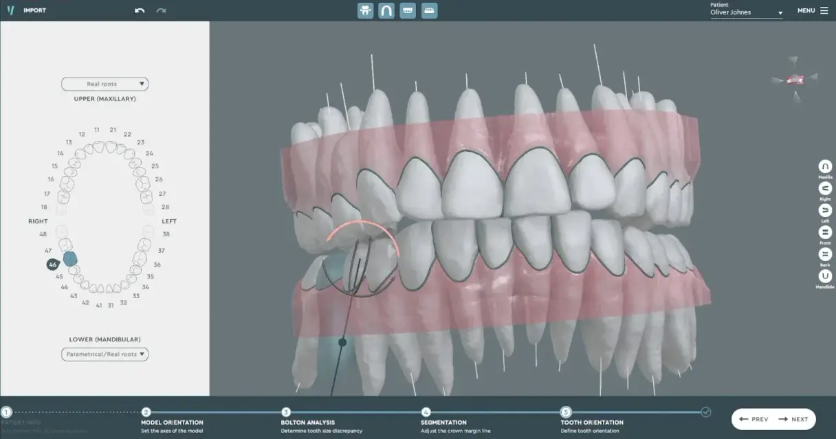
Why are parametrical roots required when real roots are available?
When real roots aren’t available, users still need to estimate what the roots look like. VISION will assist the user in estimating the real roots when they are not available. Parametrical roots are not arbitrary and can be easily adjusted to be as close to real roots as possible. If real roots are available, then this is preferable. Currently, only root analysis is available, though we plan to release bone analysis soon.
Is it possible to change the axis and center of resistance of a tooth with a real root?
Yes, this can be configured in the VISION settings menu.
Does VISION have both root and bone analysis?
Currently, only root analysis is available, though we plan to release bone analysis soon.
If overlaps between the teeth exceed IPR volume does VISION alert the user?
Yes, VISON does alert the user and is able to define the threshold.
How does a user merge CBCT DICOM files with scan STL files?
The user completes segmentation with an intra-oral scan and VISION does the rest of the work automatically.
Are users able to use PAN instead of CBCT?
Users can upload a JPEG of PAN, but it does not have volumetric data so you can review it as a reference. PAN does not have 3D information to extract any data for CBCT analysis.
To learn more about VISION and its CBCT capabilities, schedule a demonstration here.
Sign up for our newsletter!
Case Studies, Podcasts, Ebooks, Events, Webinars, Company news, and more...
By subscribing you agree to the Terms of Use and Privacy Policy.


